
Positron emission tomography (PET) is a nuclear imaging method that is used to produce 3-dimensional molecular images of the inside of the body. PET images provide detailed information about metabolic/cellular activities occurring in the body, which is needed to help diagnose and/or guide treatments for certain diseases. Radioactive PET tracers that target a disease process of interest (e.g. inflammation, calcification, fibrosis activity) are injected via a plastic tube in the arm and accumulate in areas of the body where that process is active, emitting radiation that can be detected and localised by the PET scanner. The scanner can then create a map of disease activity in the body. PET scans are typically combined with computed tomography (CT), or less commonly magnetic resonance imaging (MRI), scans for more precise anatomical localization. PET-CT is used in some patients with IMID-CVD to detect inflammation in the blood vessels and the heart muscle. However, there is a need to standardise imaging protocols and methods used to interpret PET scans in patients with IMID-CVD, to improve the consistency and reproducibility of findings across different hospital sites.
18F-fluorodeoxyglucose (FDG) is the most widely used PET tracer in clinical medicine for a variety of indications. However, as a glucose analogue, its use in the cardiovascular system is hindered by a lack of cellular specificity and high background physiological update in the myocardium. More selective PET radiotracers have emerged for cardiovascular imaging including the re-purposing of 68Ga-DOTA(TOC/NOC/TATE) tracers targeting the somatostatin receptor subtype-2 (SSTR2) expressed on macrophages (inflammation), 18F-sodium fluoride targeting developing hydroxyapatite (calcification activity) and 68Ga-FAPI which binds activated fibroblasts (fibrosis activity). PET-CT can therefore characterise the key pathological processes driving the development of cardiovascular disease in patients with IMIDs, affecting the large vessels, coronary arteries and myocardium, and potentially provide incremental diagnostic and prognostic data. Cardiovascular applications of these novel radiotracers have been pioneered by several of our Workstream Leads and will be further refined through Partnership activities.
The UK CARDIO-IMID Partnership, seeks to i) develop standardised PET-CT protocols and diagnostic criteria in IMID-CVD; ii) improve access to regional PET/CT services; iii) support collaborative multi-centre PET/CT trials; and iv) explore the application of newer PET radiotracers that are more specifically targeted to inflammation and other disease processes than those used in current clinical practice. These aims will be achieved in part by mapping the landscape of PET-CT facilities, radiopharmaceutical availability, and clinical practices across our UK Partnership sites via data collected as part of the Registry Study and ancillary surveys. Additional activities of the workstream will seek to build new cross-discipline PET/CT research initiatives to help tackle key priorities identified by the wider CARDIO-IMID Partnership.
This work and platform will strengthen partnerships between cardiovascular imaging experts and physicists and diagnostic industry partners and offer new opportunities for developing imaging biomarkers. This initiative will provide a robust platform through which to develop and utilise new and innovative methodologies, techniques, and technologies.


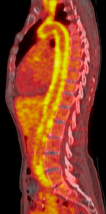
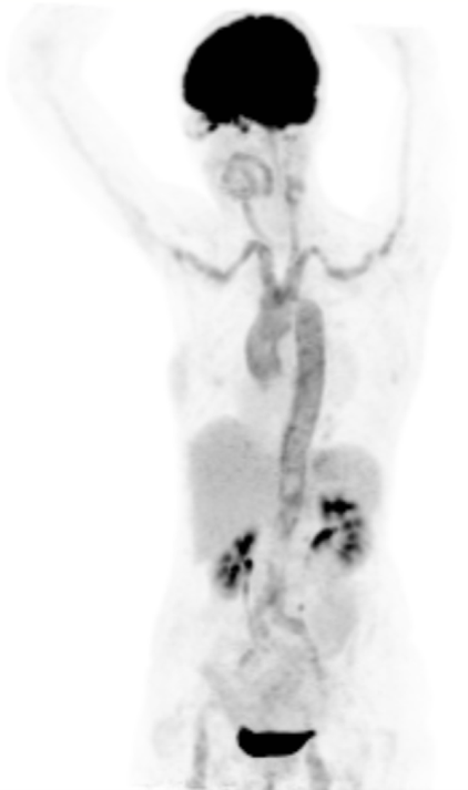
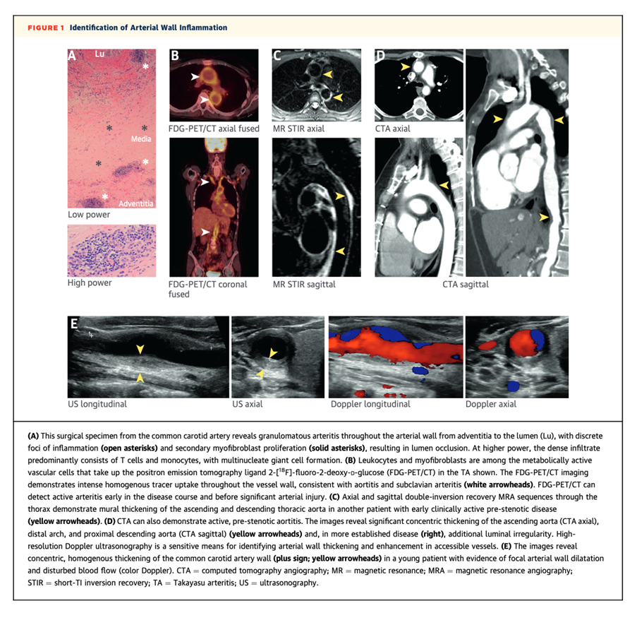
Representative 18F-FDG PET images showing active inflammation of the major blood vessels in the body due to large vessel vasculitis.


